Der Atlas enthält elektronenmikroskopische Aufnahmen vom Menschen und von Säugetieren in sehr hoher Auflösung. Tabellen und Bildübersichten ermöglichen einen schnellen Zugriff auf Abbildungen zu den Themenbereichen: Zelle, Zellkern, Zellorganellen und Cytosolstrukturen, Zelloberfläche, Zellkontakte, Cytoskelett, Extrazellularraum, Gewebe,
Epithelgewebe, Binde- und Stützgewebe, Muskelgewebe, Nervengewebe, Organe,
Blut und Lymphorgane, Haut und Sinnesorgane, Gastrointestinaltrakt, Respirationstrakt, Urogenitaltrakt, endokrine und exokrine Drüsen.
Ergänzt werden die Abbildungen durch Lehrtexte in allgemeinverständlicher Form und ein Vokabular der mikroskopischen Anatomie. Über die Präparationstechniken Transmissionselektronenmikroskopie
und Rasterelektronenmikroskopie wird ebenfalls informiert.


Schwarz-weiß-Abbildung
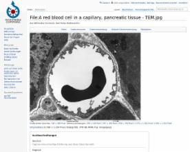
Transmission electron microscope image of a thin section cut through the pancreas(mammalian). This image shows a capillary within the pancreatic tissue(acinar cells in this image). Note the abundance of rough endoplasmic reticulum in the acinar cells. There is a red blood cell within the capillary. The capillary lining consists of long, thin endothelial cells, connected by tight junctions. The image shows fenestration of these endothelial cells. The image also shows synaptic vesicles in the neuron(nerve cell) next to the capillary.
public domain
http://en.wikipedia.org/wiki/Public_domain