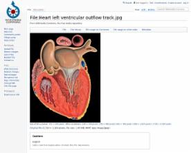
Farbdarstellung
Heart left ventricular outflow track
CC BY Patrick J. Lynch, medical illustrator 2.5
Farbgrafik
1: Blutfluss, 2: Klappe. Querschnittsdiagramm einer Vene mit Klappen.
(CC BY-derivative work: Mouagip (talk) SA 3.0
Schwarz-weiß-Darstellung
CC BY-Peter Forster SA 3.0
http://creativecommons.org/licenses/by-sa/3.0/deed.en
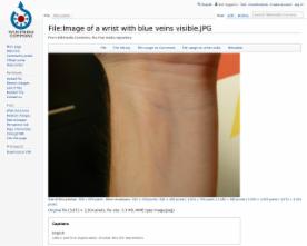
Farbfoto
An image of a wrist The blue is caused by the lack of oxygen binding to haemoglobin
CC BY-ARBAY SA 3.0
http://creativecommons.org/licenses/by-sa/3.0/deed.en
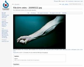
Farbfoto
CC BY Colin Davis from Chicago, United States. 2.0
http://creativecommons.org/licenses/by/2.0/deed.en
Farbfoto eines Exponats
Venengeflechte am Wirbelkanal, Wachsmodell, Paris, um 1900
CC BY User:Mattes 2.0 DE
http://creativecommons.org/licenses/by/2.0/de/deed.en
Farbabbildung
Aderlass; Gemälde des britischen Malers James Gillray um 1805, London, Victoria und Albert Museum
public domain
http://en.wikipedia.org/wiki/Public_domain
Schwarz-weiß-Abbildung
Transmission electron microscope image of a thin section cut through the pancreas(mammalian). This image shows a capillary within the pancreatic tissue(acinar cells in this image). Note the abundance of rough endoplasmic reticulum in the acinar cells. There is a red blood cell within the capillary. The capillary lining consists of long, thin endothelial cells, connected by tight junctions. The image shows fenestration of these endothelial cells. The image also shows synaptic vesicles in the neuron(nerve cell) next to the capillary.
public domain

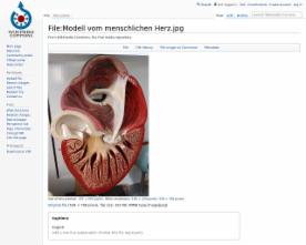






Farbfoto von einem Modell des menschlichen Herzens
CC BY-David Ludwig SA 3.0)
http://creativecommons.org/licenses/by-sa/3.0/deed.en