Farbfoto eines Exponats
Venengeflechte am Wirbelkanal, Wachsmodell, Paris, um 1900
CC BY User:Mattes 2.0 DE
http://creativecommons.org/licenses/by/2.0/de/deed.en
Farbgrafik
CC BY-A-kreislauf01.jpg: Jörg Rittmeister
derivative work: Vezixig
SA 2.5
http://creativecommons.org/licenses/by-sa/2.5/deed.de
Farbabbildung
Aderlass; Gemälde des britischen Malers James Gillray um 1805, London, Victoria und Albert Museum
public domain
http://en.wikipedia.org/wiki/Public_domain
Farbabbildung
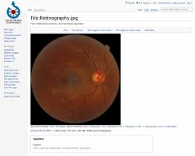
Self made ophtalmogram of the retina of the right eye. It shows the optic disc as a bright area on the right (nasal side) where blood vessels converge. The spot to the left (temporal side) of the centre is the macula. The grey, more diffuse spot in the centre is a shadow artifact.
(CC BY-Ske. SA 3.0
https://creativecommons.org/licenses/by-sa/3.0/deed.en
Farbabbildung
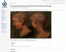
Superficial blood vessels of the head and neck. Coloured mezzotint by J. F. Gautier D'Agoty, 1748, Wellcome Library
public domain
http://commons.wikimedia.org/wiki/Public_domain#Material_in_the_public_…
Farbgrafik
Schema der Koronargefäße (Ansicht etwa von der linken Schulter). Im Vordergrund die linke, hinten die rechte Koronararterie
CC BY Patrick J. Lynch, medical illustrator 2.5
http://creativecommons.org/licenses/by/2.5/deed.en
Farbdarstellung
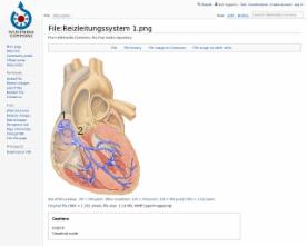
Schema des Herzens mit Erregungsleitungssystem in blau. (1) Sinusknoten, (2) AV-Knoten
CC BY J. Heuser 2.5
http://creativecommons.org/licenses/by/2.5/deed.en


Farbfoto eines Modells
CC BY GreenFlames09 2.0
http://creativecommons.org/licenses/by/2.0/deed.de